Please login in order to download photos in full size
If you are not registered, please register for free: www.Free-Photos.biz/register
Please note to download premium images you also need to join as a free member..
You can also save the photos without the registration - but only in small and average sizes, and some of them will have the site's watermark. Please simply click your right mouse button and save the image.
Please login in order to like photos
If you are not registered, please register for free:
Sorry, non-members can download up to 100 full-size photos per month.
It looks like you have used up your limit.
Free members can download an unlimited number of full-size photos - including the premium free photos.
Join as a member today for FREE! - and download the images without limitations:
www.Free-Photos.biz/membership.php
You can also save the images without the membership - but only in small and average sizes, and some of them may have the site's watermark. Please simply click your right mouse button and save the image.
|
Download top free photographs!
This photo was viewed 8 times and was downloaded in full size 0 times.
This photo was liked 0 times


Source page: | http://commons.wikimedia.org/wiki/File:Schleiden;_cellular_tissue_of_plants_Wellcome_M0010608.jpg |
|---|
Summaryedit
| Title |
Schleiden; cellular tissue of plants |
||
| Description |
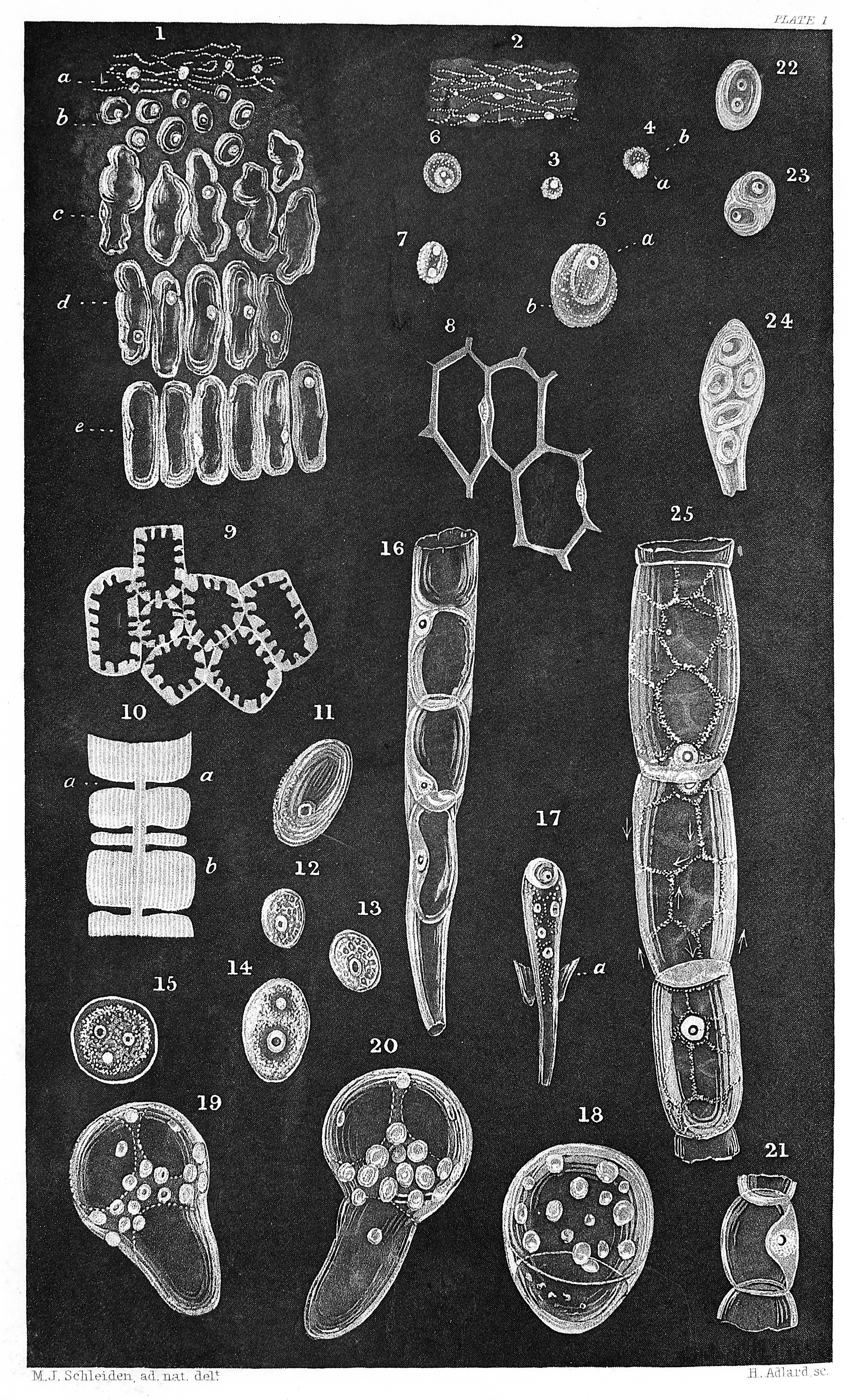
Appended to Schwann's book in the same volume; Contributions to Phytogenesis, translated from the German of Dr M. J. Schleiden, Professor of Botany at the University of Jena. Plate I Fig. 1. Cellular tissue from the embryo-sac of Chamoedorea Scheideana in the act of formation. a. The inner-most mass, consisting of bum with intermingled mucous granules and cytoblasts. b. Newly formed cells, still soluble in distilled water. c-e. Further development of the cells, which, with the exception of the cytoblasts, may still coalesce, under slight pressure, into an amorphous mass. 2. the formative substance from fig. 1, a, more highly magnified, bum, mucous granules, nuclei of the cytoblasts, and cytoblasts. 3. a single and as yet free cytoblast, still more highly magnified. 4. A cytoblast with the cell forming upon it. 5. The same, more highly magnified. 6. The same. The cytoblast here exhibits two nuclei, and is delineated in 7, isolated after the destruction of the cell by pressure. 8. the same cellular tissue in a higher degree of development that that represented by fig. 1, e. The contiguous cell-walls have already united. By making a transverse section, it may be distinctly perceived the that cytoblast is enclosed in the cell-wall. 9. Cells from a delicate transverse sectionof the almost matured albumen. 10. Common partition-wall between two cells from fig. 9, under a higher magnifying power. The stratiform depositions may be ovserved at b, and the porous canals produced by their local failure at a. I could distinctly enumerate from nine to twelve layers which had been deposited within fourteen days. 11. a sporule from Rhizina laevigata Fries, with the cytoblast. 12, 13, 14. Different cytoblasts from the embryo-sac of Pimelea drupacea before the appearance of cells. 15. A young cell with its cytoblast, from the same. The latter in this instance presents the unusual number of three nucleoli. 16. A portion of the embryonal end of the pollen-tube projecting from the ovulum in Orchis Morio, within which, towards the upper part, cells have been already developed. At the lower part, the original pollen-tube may still be distinguished. The almost globular cytoblasts are, in this instance, distinctly enclosed in the cell-wall. 17. Embryonal end of the pollen-tube from Linum pallescens, together with an appended lobule of the embryo-sac (a). The process of the formation of cells is commencing. Above, a young cell with its cytoblast is already perceptible, beneath this several cell-nuclei are seen floating free. 18, 19, 20. Commencing germination in the sporules of Marchantia polymorpha. Compare the text, p. 248. 21. Portions of the pollen-tube which have become cellular, from Orchis latifolia, in the highest stage of development; the investment of the pollen-tube is no longer perceptible. the cytoblast is enclosed in the cell-wall, just as in fig. 16. 22 and 23. Two isolated cells from the terminal shoot (punctum vegetationis, Wolff) of Gasteria racemosa; 22 exhibitis two free cytoblasts; 23, two newly-formed cells within the original cell. Fig 24. A very young leaf of Crassula portulaca, the five cells which solely compose it being still surrounded by a parent cell. 25. Three cells from an articulated hair of potato, with a retiform current of mucus upon their walls. In the central cell the direction of the currents is partially indicated by arrows. Rare Books |
||
| Credit line |
|
||
| References |
|
||
| Source/Photographer |
https://wellcomeimages.org/indexplus/obf_images/55/5c/5207ca96c667b2f07440c28cd4b1.jpg
|
||
| Other versions |
  |
Licensingedit
| This file is licensed under the Creative Commons Attribution 4.0 International license. | ||
|
| EXIF data: | |
| File name | schleiden__cellular_tissue_of_plants_wellcome_m0010608.jpg |
|---|---|
| Size, Mbytes | 4.091021484375 |
| Mime type | image/jpeg |
While the copyright and licensing information supplied for each photo is believed to be accurate, Free-Photos.biz does not provide any warranty regarding the copyright status or correctness of licensing terms. If you decide to reuse the images from Free-Photos.biz, you should verify the copyright status of each image just as you would when obtaining images from other sources.
The use of depictions of living or deceased persons may be restricted in some jurisdictions by laws regarding personality rights. Such images are exhibited at Free-Photos.biz as works of art that serve higher artistic interests.
PRIVACY POLICY
By registering your account and/or by subscribing to new and newly rated photographs you agree we may send you the links to photos and we may occasionally share other information with you.
We do NOT disclose your personal data.